


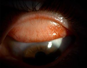
 Closed until 9am
This is the only clinic technique that can provide information on the state of your meibomian glands. By using infrared technology, the structure of the glands can be looked at accurately with no interference from the surrounding anatomy.
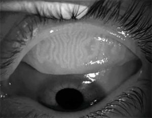
We have around 20-30 glands on the lower eye lids and 30-40 on the upper eye lids. The imaging allows us to see whether the glands are structurally normal, dilated, shortened or atrophied from chronic obstruction.
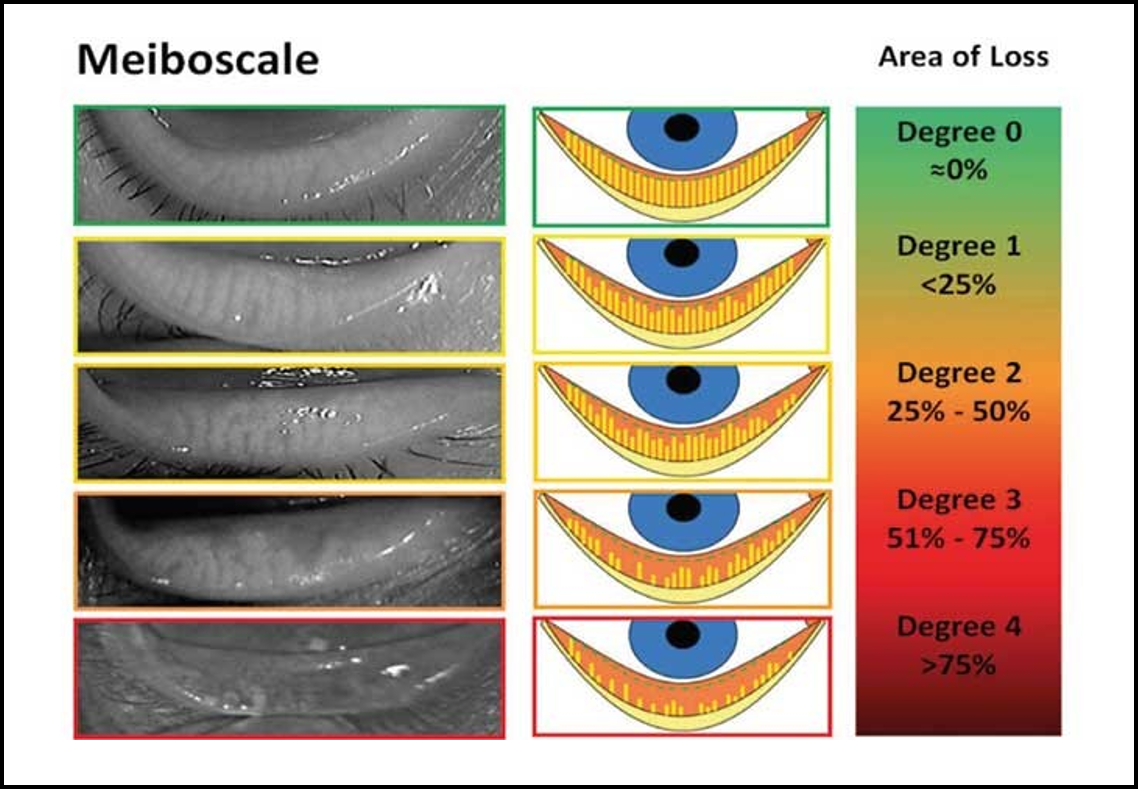
With Meibography we can grade the level of meibomian gland dysfunction on each eyelid:
Once we can visualise the anatomy of the glands, diagnostic meibomian gland expression is performed to determine the quality of ‘meibum’ that is produced.