


 Open today until 7pm
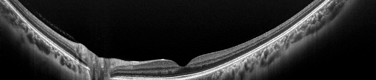
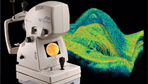 Optical Coherence Tomography (OCT) is a non-invasive, non contact imaging technique that produces high resolution images of the retina. It is similar to ultrasound except it uses light rather than sound waves to produce a much higher resolution image.
OCT has been shown to be clinically useful for evaluating eye disease like macular holes, macular edema, macular degeneration, epiretinal membranes and glaucoma. It is used to visualize nearly transparent structures of the eye, examine the extent of retinal defects or abnormalities cause by trauma, perform detailed measurements to determine the specific cause and develop treatment plans for conditions and monitor outcomes of treatment procedures over time.
Dr. Saari believes that knowing the fine details of your retina before any conditions are detected is crucial when evaluating newly detected disease. This is why we perform baseline OCT images with photography on all patients during their first examination at no additional charge.
In some cases dilation drops are not necessary but we do advise patients to be prepared for the typical effects of dilation which include blur at near and sensitivity to bright lights after this procedure.
OCT is not an insured service through provincial health insurance (OHIP) for any patient when performed by an optometrist and in limited cases when performed by an ophthalmologist.